

Mercury Causes Brain Damage (Autism™)
Thimerosal is mercury.

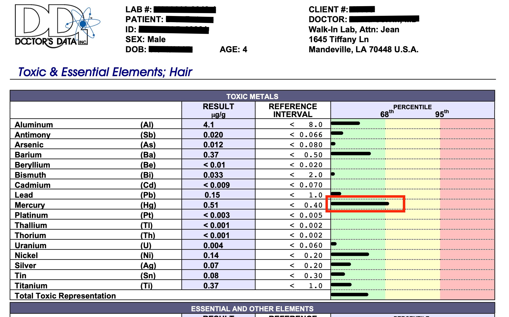
The lab results for toxic heavy metals using a hair sample from a child injected with thimerosal with severe autism are shown below:
How Mercury Causes Brain Neuron Degeneration
Mercury has long been know to be a potent neurotoxic substance, whether it is inhaled or consumed in a diet as a food contaminant. Over the past 15 years medical research laboratories have established that dental amalgam tooth fillings are a major contributor to mercury-body burden.
In 1997, a team of research scientists demonstrated that mercury vapor inhalation by animals, produced a molecular lesion in brain protein metabolism, which was similar to a lesion seen in 80 percent of Alzheimer's diseased brains. Recently completed experiments by scientists at the University of Calgary's Faculty of Medicine now reveal, with direct visual evidence from brain neuron tissue cultures, how mercury ions actually alter the cell membrane structure of developing neurons.
To better understand Mercury's effect on the brain, let us first illustrate what brain neurons look like and how they grow.
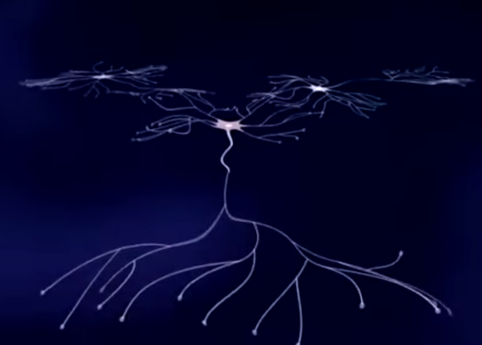
In this illustration, we see three brain neurons growing in a tissue culture, each with a central cell body and numerous neurite processes1.

At the end of each neurite is a growth cone where structural proteins are assembled to form the cell membrane.

Two principal proteins involved in growth cone function are actin2 which is responsible for the pulsating motion, and tubulin3, a major structural component of the neurite membrane.
During normal cell growth, tubulin molecules link together end-to-end to form microtubules, which surround neurofibrils, another structural protein component of the neuronal axon.

Shown here is the neurite of a live neuron isolated from snail brain tissue, displaying linear growth due to growth cone activity.

It is important to note that growth cones in all animal species, ranging from snails to humans, have identical structural and behavioral characteristics, and use proteins of virtually identical composition.
In this experiment, neurons also isolated from snail brain tissue were grown in culture for several days, after which very low concentrations of mercury were added to the culture medium for 20 minutes.
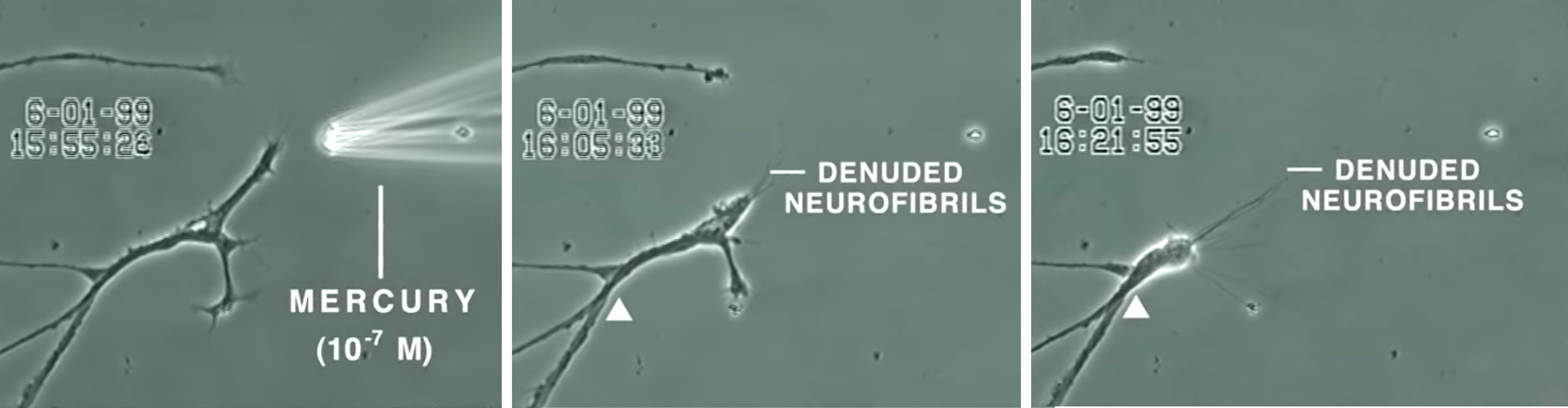
Over the next 30 minutes, the neurite membrane underwent rapid degeneration, leaving behind the denuded neurofibrils4 seen in the illustration above.
In contrast, other heavy metals added at this same concentration, such as aluminum, lead, cadmium, and manganese, did not produce this effect.
To understand how mercury causes this degeneration, let us return to our illustration. As mentioned before, tubulin proteins link together during normal cell growth to form the microtubules which support the neurite structure.

When mercury ions are introduced into the culture medium, they infiltrate the cell and bind themselves to newly synthesized tubulin molecules. More specifically, the mercury ions attach themselves to the binding site reserved for guanosine triphosphate, or GTP, on the beta subunit of the affected tubulin molecules. Since bound GTP normally provides the energy which allows tubulin molecules to attach to one another, mercury ions bound to these sites prevent tubulin proteins from linking together.
Consequently, the neurite's microtubules begin to disassemble into free tubulin molecules, leaving the neurite stripped of its supporting structure. Ultimately, both the developing neurite and its growth cone collapse, and some denuded neurofibrils form aggregates or tangles, as depicted here:

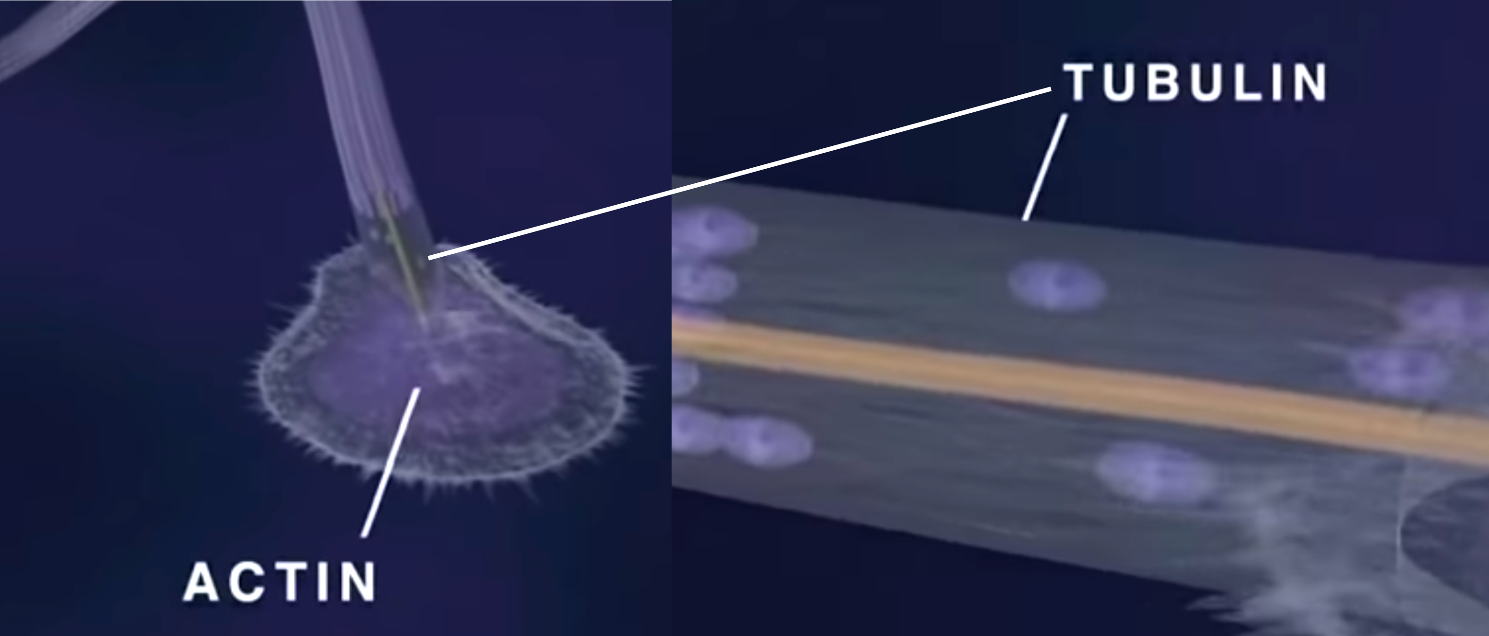
Shown here is a neurite growth cone stained specifically for tubulin and actin before and after mercury exposure:

Note that the mercury has caused disintegration of tubulin microtubule structure. These new findings reveal important visual evidence as to how mercury causes neurodegeneration. More importantly, the study provides the first direct evidence that low-level mercury exposure is indeed a precipitating factor that can initiate this neurodegenerative process within the brain.
The scientists:
F.L. Lorscheider.
C. C-W. Leong.
N. I. Syed
Dept. of Physiology and Biophysics Faculty of Medicine
University of Calgary
Autism: a novel form of mercury poisoning
Abstract
Autism is a syndrome characterized by impairments in social relatedness and communication, repetitive behaviors, abnormal movements, and sensory dysfunction. Recent epidemiological studies suggest that autism may affect 1 in 150 US children. Exposure to mercury can cause immune, sensory, neurological, motor, and behavioral dysfunctions similar to traits defining or associated with autism, and the similarities extend to neuroanatomy, neurotransmitters, and biochemistry. Thimerosal, a preservative added to many vaccines, has become a major source of mercury in children who, within their first two years, may have received a quantity of mercury that exceeds safety guidelines. A review of medical literature and US government data suggests that: (i) many cases of idiopathic autism are induced by early mercury exposure from thimerosal; (ii) this type of autism represents an unrecognized mercurial syndrome; and (iii) genetic and non-genetic factors establish a predisposition whereby thimerosal's adverse effects occur only in some children.
Source: https://pubmed.ncbi.nlm.nih.gov/11339848/
A two-phase study evaluating the relationship between Thimerosal-containing vaccine administration and the risk for an autism spectrum disorder diagnosis in the United States
Abstract
Background: Autism spectrum disorder (ASD) is defined by standardized criteria of qualitative impairments in social interaction, qualitative impairments in communication, and restricted and stereotyped patterns of behavior, interests, and activities. A significant number of children diagnosed with ASD suffer a loss of previously-acquired skills, which is suggestive of neurodegeneration or a type of progressive encephalopathy with an etiological pathogenic basis occurring after birth. To date, the etiology of ASD remains under debate, however, many studies suggest toxicity, especially from mercury (Hg), in individuals diagnosed with an ASD. The present study evaluated concerns about the toxic effects of organic-Hg exposure from Thimerosal (49.55% Hg by weight) in childhood vaccines by conducting a two-phased (hypothesis generating/hypothesis testing) study with documented exposure to varying levels of Thimerosal from vaccinations.
Source: https://pubmed.ncbi.nlm.nih.gov/24354891/
A positive association found between autism prevalence and childhood vaccination uptake across the U.S. population
Abstract
The reason for the rapid rise of autism in the United States that began in the 1990s is a mystery. Although individuals probably have a genetic predisposition to develop autism, researchers suspect that one or more environmental triggers are also needed. One of those triggers might be the battery of vaccinations that young children receive. Using regression analysis and controlling for family income and ethnicity, the relationship between the proportion of children who received the recommended vaccines by age 2 years and the prevalence of autism (AUT) or speech or language impairment (SLI) in each U.S. state from 2001 and 2007 was determined. A positive and statistically significant relationship was found: The higher the proportion of children receiving recommended vaccinations, the higher was the prevalence of AUT or SLI. A 1% increase in vaccination was associated with an additional 680 children having AUT or SLI. Neither parental behavior nor access to care affected the results, since vaccination proportions were not significantly related (statistically) to any other disability or to the number of pediatricians in a U.S. state. The results suggest that although mercury has been removed from many vaccines, other culprits may link vaccines to autism. Further study into the relationship between vaccines and autism is warranted.
Source: https://pubmed.ncbi.nlm.nih.gov/21623535/
Commentary--Controversies surrounding mercury in vaccines: autism denial as impediment to universal immunisation
Abstract
In 2004, the US Center for Disease Control (CDC) published a paper showing that there is no link between the age at which a child is vaccinated with MMR and the vaccinated children's risk of a subsequent diagnosis of autism. One of the authors, William Thompson, has now revealed that statistically significant information was deliberately omitted from the paper. Thompson first told Dr S Hooker, a researcher on autism, about the manipulation of the data. Hooker analysed the raw data from the CDC study afresh. He confirmed that the risk of autism among African American children vaccinated before the age of 2 years was 340% that of those vaccinated later.
Source: https://pubmed.ncbi.nlm.nih.gov/25377033/
Methodological issues and evidence of malfeasance in research purporting to show thimerosal in vaccines is safe
Abstract
There are over 165 studies that have focused on Thimerosal, an organic-mercury (Hg) based compound, used as a preservative in many childhood vaccines, and found it to be harmful. Of these, 16 were conducted to specifically examine the effects of Thimerosal on human infants or children with reported outcomes of death; acrodynia; poisoning; allergic reaction; malformations; auto-immune reaction; Well's syndrome; developmental delay; and neurodevelopmental disorders, including tics, speech delay, language delay, attention deficit disorder, and autism. In contrast, the United States Centers for Disease Control and Prevention states that Thimerosal is safe and there is "no relationship between [T]himerosal[-]containing vaccines and autism rates in children." This is puzzling because, in a study conducted directly by CDC epidemiologists, a 7.6-fold increased risk of autism from exposure to Thimerosal during infancy was found. The CDC's current stance that Thimerosal is safe and that there is no relationship between Thimerosal and autism is based on six specific published epidemiological studies coauthored and sponsored by the CDC. The purpose of this review is to examine these six publications and analyze possible reasons why their published outcomes are so different from the results of investigations by multiple independent research groups over the past 75+ years.
Source: https://pubmed.ncbi.nlm.nih.gov/24995277/
Abnormal measles-mumps-rubella antibodies and CNS autoimmunity in children with autism
Abstract
Autoimmunity to the central nervous system (CNS), especially to myelin basic protein (MBP), may play a causal role in autism, a neurodevelopmental disorder. Because many autistic children harbor elevated levels of measles antibodies, we conducted a serological study of measles-mumps-rubella (MMR) and MBP autoantibodies. Using serum samples of 125 autistic children and 92 control children, antibodies were assayed by ELISA or immunoblotting methods. ELISA analysis showed a significant increase in the level of MMR antibodies in autistic children. Immunoblotting analysis revealed the presence of an unusual MMR antibody in 75 of 125 (60%) autistic sera but not in control sera. This antibody specifically detected a protein of 73-75 kD of MMR. This protein band, as analyzed with monoclonal antibodies, was immunopositive for measles hemagglutinin (HA) protein but not for measles nucleoprotein and rubella or mumps viral proteins. Thus the MMR antibody in autistic sera detected measles HA protein, which is unique to the measles subunit of the vaccine. Furthermore, over 90% of MMR antibody-positive autistic sera were also positive for MBP autoantibodies, suggesting a strong association between MMR and CNS autoimmunity in autism. Stemming from this evidence, we suggest that an inappropriate antibody response to MMR, specifically the measles component thereof, might be related to pathogenesis of autism.
Source: https://pubmed.ncbi.nlm.nih.gov/12145534/
Hepatitis B vaccination of male neonates and autism diagnosis, NHIS 1997-2002
Abstract
Universal hepatitis B vaccination was recommended for U.S. newborns in 1991; however, safety findings are mixed. The association between hepatitis B vaccination of male neonates and parental report of autism diagnosis was determined. This cross-sectional study used weighted probability samples obtained from National Health Interview Survey 1997-2002 data sets. Vaccination status was determined from the vaccination record. Logistic regression was used to estimate the odds for autism diagnosis associated with neonatal hepatitis B vaccination among boys age 3-17 years, born before 1999, adjusted for race, maternal education, and two-parent household. Boys vaccinated as neonates had threefold greater odds for autism diagnosis compared to boys never vaccinated or vaccinated after the first month of life. Non-Hispanic white boys were 64% less likely to have autism diagnosis relative to nonwhite boys. Findings suggest that U.S. male neonates vaccinated with the hepatitis B vaccine prior to 1999 (from vaccination record) had a threefold higher risk for parental report of autism diagnosis compared to boys not vaccinated as neonates during that same time period. Nonwhite boys bore a greater risk.
Source: https://pubmed.ncbi.nlm.nih.gov/21058170/
Do aluminum vaccine adjuvants contribute to the rising prevalence of autism?
Abstract
Autism spectrum disorders (ASD) are serious multisystem developmental disorders and an urgent global public health concern. Dysfunctional immunity and impaired brain function are core deficits in ASD. Aluminum (Al), the most commonly used vaccine adjuvant, is a demonstrated neurotoxin and a strong immune stimulator. Hence, adjuvant Al has the potential to induce neuroimmune disorders. When assessing adjuvant toxicity in children, two key points ought to be considered: (i) children should not be viewed as "small adults" as their unique physiology makes them much more vulnerable to toxic insults; and (ii) if exposure to Al from only few vaccines can lead to cognitive impairment and autoimmunity in adults, is it unreasonable to question whether the current pediatric schedules, often containing 18 Al adjuvanted vaccines, are safe for children? By applying Hill's criteria for establishing causality between exposure and outcome we investigated whether exposure to Al from vaccines could be contributing to the rise in ASD prevalence in the Western world. Our results show that: (i) children from countries with the highest ASD prevalence appear to have the highest exposure to Al from vaccines; (ii) the increase in exposure to Al adjuvants significantly correlates with the increase in ASD prevalence in the United States observed over the last two decades (Pearson r=0.92, p<0.0001); and (iii) a significant correlation exists between the amounts of Al administered to preschool children and the current prevalence of ASD in seven Western countries, particularly at 3-4 months of age (Pearson r=0.89-0.94, p=0.0018-0.0248). The application of the Hill's criteria to these data indicates that the correlation between Al in vaccines and ASD may be causal. Because children represent a fraction of the population most at risk for complications following exposure to Al, a more rigorous evaluation of Al adjuvant safety seems warranted.
Source: https://pubmed.ncbi.nlm.nih.gov/22099159/
What is regressive autism and why does it occur? Is it the consequence of multi-systemic dysfunction affecting the elimination of heavy metals and the ability to regulate neural temperature?
Abstract
There is a compelling argument that the occurrence of regressive autism is attributable to genetic and chromosomal abnormalities, arising from the overuse of vaccines, which subsequently affects the stability and function of the autonomic nervous system and physiological systems. That sense perception is linked to the autonomic nervous system and the function of the physiological systems enables us to examine the significance of autistic symptoms from a systemic perspective. Failure of the excretory system influences elimination of heavy metals and facilitates their accumulation and subsequent manifestation as neurotoxins: the long-term consequences of which would lead to neurodegeneration, cognitive and developmental problems. It may also influence regulation of neural hyperthermia. This article explores the issues and concludes that sensory dysfunction and systemic failure, manifested as autism, is the inevitable consequence arising from subtle DNA alteration and consequently from the overuse of vaccines.
Source: https://pubmed.ncbi.nlm.nih.gov/22666668/
A case series of children with apparent mercury toxic encephalopathies manifesting with clinical symptoms of regressive autistic disorders
Abstract
Impairments in social relatedness and communication, repetitive behaviors, and stereotypic abnormal movement patterns characterize autism spectrum disorders (ASDs). It is clear that while genetic factors are important to the pathogenesis of ASDs, mercury exposure can induce immune, sensory, neurological, motor, and behavioral dysfunctions similar to traits defining or associated with ASDs. The Institutional Review Board of the Institute for Chronic Illnesses (Office for Human Research Protections, U.S. Department of Health and Human Services, IRB number IRB00005375) approved the present study. A case series of nine patients who presented to the Genetic Centers of America for a genetic/developmental evaluation are discussed. Eight of nine patients (one patient was found to have an ASD due to Rett's syndrome) (a) had regressive ASDs; (b) had elevated levels of androgens; (c) excreted significant amounts of mercury post chelation challenge; (d) had biochemical evidence of decreased function in their glutathione pathways; (e) had no known significant mercury exposure except from Thimerosal-containing vaccines/Rho(D)-immune globulin preparations; and (f) had alternate causes for their regressive ASDs ruled out. There was a significant dose-response relationship between the severity of the regressive ASDs observed and the total mercury dose children received from Thimerosal-containing vaccines/Rho (D)-immune globulin preparations. Based upon differential diagnoses, 8 of 9 patients examined were exposed to significant mercury from Thimerosal-containing biologic/vaccine preparations during their fetal/infant developmental periods, and subsequently, between 12 and 24 mo of age, these previously normally developing children suffered mercury toxic encephalopathies that manifested with clinical symptoms consistent with regressive ASDs. Evidence for mercury intoxication should be considered in the differential diagnosis as contributing to some regressive ASDs.
Source: https://pubmed.ncbi.nlm.nih.gov/17454560/
A comprehensive review of mercury provoked autism
Abstract
Emerging evidence supports the theory that some autism spectrum disorders (ASDs) may result from a combination of genetic/biochemical susceptibility, specifically a reduced ability to excrete mercury (Hg), and exposure to Hg at critical developmental periods. Elemental/inorganic Hg is released into the air/water where it becomes methylated and accumulates in animal tissues. The US population is primarily exposed to methyl-Hg by fish consumption. In addition, many pharmaceuticals have been, and some continue to be, a ubiquitous source of danger because they contain mercurials. Mercurials may be found in drugs for the eye, ear, nose, throat, and skin; in bleaching creams; as preservatives in cosmetics, tooth pastes, lens solutions, vaccines, allergy test and immunotherapy solutions; in antiseptics, disinfectants, and contraceptives; in fungicides and herbicides; in dental fillings and thermometers; and many other products. Hg has been found to cause immune, sensory, neurological, motor, and behavioural dysfunctions similar to traits defining/associated with ASDs, and that these similarities extend to neuroanatomy, neurotransmitters, and biochemistry. Furthermore, a review of molecular mechanisms indicates that Hg exposure can induce death, disorganization and/or damage to selected neurons in the brain similar to that seen in recent ASD brain pathology studies, and this alteration may likely produce the symptoms by which ASDs are diagnosed. Finally, a review of treatments suggests that ASD patients who undergo protocols to reduce Hg and/or its effects show significant clinical improvements in some cases. In conclusion, the overwhelming preponderance of the evidence favours acceptance that Hg exposure is capable of causing some ASDs.
Source: https://pubmed.ncbi.nlm.nih.gov/19106436/
Thimerosal exposure and the role of sulfation chemistry and thiol availability in autism
Abstract
Autism spectrum disorder (ASD) is a neurological disorder in which a significant number of the children experience a developmental regression characterized by a loss of previously acquired skills and abilities. Typically reported are losses of verbal, nonverbal, and social abilities. Several recent studies suggest that children diagnosed with an ASD have abnormal sulfation chemistry, limited thiol availability, and decreased glutathione (GSH) reserve capacity, resulting in a compromised oxidation/reduction (redox) and detoxification capacity. Research indicates that the availability of thiols, particularly GSH, can influence the effects of thimerosal (TM) and other mercury (Hg) compounds. TM is an organomercurial compound (49.55% Hg by weight) that has been, and continues to be, used as a preservative in many childhood vaccines, particularly in developing countries. Thiol-modulating mechanisms affecting the cytotoxicity of TM have been identified. Importantly, the emergence of ASD symptoms post-6 months of age temporally follows the administration of many childhood vaccines. The purpose of the present critical review is provide mechanistic insight regarding how limited thiol availability, abnormal sulfation chemistry, and decreased GSH reserve capacity in children with an ASD could make them more susceptible to the toxic effects of TM routinely administered as part of mandated childhood immunization schedules.
Source: https://pubmed.ncbi.nlm.nih.gov/23965928/
B-lymphocytes from a population of children with autism spectrum disorder and their unaffected siblings exhibit hypersensitivity to thimerosal
Abstract
The role of thimerosal containing vaccines in the development of autism spectrum disorder (ASD) has been an area of intense debate, as has the presence of mercury dental amalgams and fish ingestion by pregnant mothers. We studied the effects of thimerosal on cell proliferation and mitochondrial function from B-lymphocytes taken from individuals with autism, their nonautistic twins, and their nontwin siblings. Eleven families were examined and compared to matched controls. B-cells were grown with increasing levels of thimerosal, and various assays (LDH, XTT, DCFH, etc.) were performed to examine the effects on cellular proliferation and mitochondrial function. A subpopulation of eight individuals (4 ASD, 2 twins, and 2 siblings) from four of the families showed thimerosal hypersensitivity, whereas none of the control individuals displayed this response. The thimerosal concentration required to inhibit cell proliferation in these individuals was only 40% of controls. Cells hypersensitive to thimerosal also had higher levels of oxidative stress markers, protein carbonyls, and oxidant generation. This suggests certain individuals with a mild mitochondrial defect may be highly susceptible to mitochondrial specific toxins like the vaccine preservative thimerosal.
Source: https://pubmed.ncbi.nlm.nih.gov/23843785/
Theoretical aspects of autism: causes--a review
Abstract
Autism, a member of the pervasive developmental disorders (PDDs), has been increasing dramatically since its description by Leo Kanner in 1943. First estimated to occur in 4 to 5 per 10,000 children, the incidence of autism is now 1 per 110 in the United States, and 1 per 64 in the United Kingdom, with similar incidences throughout the world. Searching information from 1943 to the present in PubMed and Ovid Medline databases, this review summarizes results that correlate the timing of changes in incidence with environmental changes. Autism could result from more than one cause, with different manifestations in different individuals that share common symptoms. Documented causes of autism include genetic mutations and/or deletions, viral infections, and encephalitis following vaccination. Therefore, autism is the result of genetic defects and/or inflammation of the brain. The inflammation could be caused by a defective placenta, immature blood-brain barrier, the immune response of the mother to infection while pregnant, a premature birth, encephalitis in the child after birth, or a toxic environment.
Source: https://pubmed.ncbi.nlm.nih.gov/21299355/
A prospective study of thimerosal-containing Rho(D)-immune globulin administration as a risk factor for autistic disorders
Abstract
Background: This study evaluated the relationship between prenatal mercury exposure from thimerosal (49.55% mercury by weight)-containing Rho(D)-immune globulins (TCRs) and autism spectrum disorders (ASDs).
Source: https://pubmed.ncbi.nlm.nih.gov/17674242/
Hypothesis: conjugate vaccines may predispose children to autism spectrum disorders
Abstract
The first conjugate vaccine was approved for use in the US in 1988 to protect infants and young children against the capsular bacteria Haemophilus influenzae type b (Hib). Since its introduction in the US, this vaccine has been approved in most developed countries, including Denmark and Israel where the vaccine was added to their national vaccine programs in 1993 and 1994, respectively. There have been marked increases in the reported prevalence of autism spectrum disorders (ASDs) among children in the US beginning with birth cohorts in the late 1980s and in Denmark and Israel starting approximately 4-5 years later. Although these increases may partly reflect ascertainment biases, an exogenous trigger could explain a significant portion of the reported increases in ASDs. It is hypothesized here that the introduction of the Hib conjugate vaccine in the US in 1988 and its subsequent introduction in Denmark and Israel could explain a substantial portion of the initial increases in ASDs in those countries. The continuation of the trend toward increased rates of ASDs could be further explained by increased usage of the vaccine, a change in 1990 in the recommended age of vaccination in the US from 15 to 2 months, increased immunogenicity of the vaccine through changes in its carrier protein, and the subsequent introduction of the conjugate vaccine for Streptococcus pneumoniae. Although conjugate vaccines have been highly effective in protecting infants and young children from the significant morbidity and mortality caused by Hib and S. pneumoniae, the potential effects of conjugate vaccines on neural development merit close examination. Conjugate vaccines fundamentally change the manner in which the immune systems of infants and young children function by deviating their immune responses to the targeted carbohydrate antigens from a state of hypo-responsiveness to a robust B2 B cell mediated response. This period of hypo-responsiveness to carbohydrate antigens coincides with the intense myelination process in infants and young children, and conjugate vaccines may have disrupted evolutionary forces that favored early brain development over the need to protect infants and young children from capsular bacteria.
Source: https://pubmed.ncbi.nlm.nih.gov/21993250/
The potential importance of steroids in the treatment of autistic spectrum disorders and other disorders involving mercury toxicity
Abstract
Autism is a neurodevelopmental disorder that according to the Centers for Disease Control and Prevention (CDC) affects 1 in 150 children in the United States. Autism is characterized by impairments in social relatedness and communication, repetitive behaviors, abnormal movements, and sensory dysfunction. Recently emerging evidence suggests that mercury, especially from childhood vaccines, appears to be a factor in the development of the autistic disorders, and that autistic children have higher than normal body-burdens of mercury. In considering mercury toxicity, it has previously been shown that testosterone significantly potentates mercury toxicity, whereas estrogen is protective. Examination of autistic children has shown that the severity of autistic disorders correlates with the amount of testosterone present in the amniotic fluid, and an examination of a case-series of autistic children has shown that some have plasma testosterone levels that were significantly elevated in comparison neurotypical control children. A review of some of the current biomedical therapies for autistics, such as glutathione and cysteine, chelation, secretin, and growth hormone, suggests that they may in fact lower testosterone levels. We put forward the medical hypothesis that autistic disorders, in fact, represents a form of testosterone mercury toxicity, and based upon this observation, one can design novel treatments for autistics directed towards higher testosterone levels in autistic children. We suggest a series of experiments that need to be conducted in order to evaluate the exact mechanisms for mercury-testosterone toxicity, and various types of clinical manipulations that may be employed to control testosterone levels. It is hoped by devising therapies that address the steroid hormone pathways, in addition to the current treatments that successful lower heavy metal body-burdens of mercury, will work synergistically to improve clinical outcomes. In light of the fact that there are a number of other diseases that may have a chronic mercury toxicity component, such as Alzheimer's disease, heart disease, obesity, ALS, asthma, and other various forms of autoimmune disorders, it is imperative that further research should be conducted to understand mercury-testosterone toxicity.
Source: https://pubmed.ncbi.nlm.nih.gov/15780490/
Reduced levels of mercury in first baby haircuts of autistic children
Abstract
Reported rates of autism have increased sharply in the United States and the United Kingdom. One possible factor underlying these increases is increased exposure to mercury through thimerosal-containing vaccines, but vaccine exposures need to be evaluated in the context of cumulative exposures during gestation and early infancy. Differential rates of postnatal mercury elimination may explain why similar gestational and infant exposures produce variable neurological effects. First baby haircut samples were obtained from 94 children diagnosed with autism using Diagnostic and Statistical Manual of Mental Disorders, 4th edition (DSM IV) criteria and 45 age- and gender-matched controls. Information on diet, dental amalgam fillings, vaccine history, Rho D immunoglobulin administration, and autism symptom severity was collected through a maternal survey questionnaire and clinical observation. Hair mercury levels in the autistic group were 0.47 ppm versus 3.63 ppm in controls, a significant difference. The mothers in the autistic group had significantly higher levels of mercury exposure through Rho D immunoglobulin injections and amalgam fillings than control mothers. Within the autistic group, hair mercury levels varied significantly across mildly, moderately, and severely autistic children, with mean group levels of 0.79, 0.46, and 0.21 ppm, respectively. Hair mercury levels among controls were significantly correlated with the number of the mothers' amalgam fillings and their fish consumption as well as exposure to mercury through childhood vaccines, correlations that were absent in the autistic group. Hair excretion patterns among autistic infants were significantly reduced relative to control. These data cast doubt on the efficacy of traditional hair analysis as a measure of total mercury exposure in a subset of the population. In light of the biological plausibility of mercury's role in neurodevelopmental disorders, the present study provides further insight into one possible mechanism by which early mercury exposures could increase the risk of autism.
Source: https://pubmed.ncbi.nlm.nih.gov/12933322/
Cultured lymphocytes from autistic children and non-autistic siblings up-regulate heat shock protein RNA in response to thimerosal challenge
Abstract
There are reports suggesting that some autistic children are unable to mount an adequate response following exposure to environmental toxins. This potential deficit, coupled with the similarity in clinical presentations of autism and some heavy metal toxicities, has led to the suggestion that heavy metal poisoning might play a role in the etiology of autism in uniquely susceptible individuals. Thimerosal, an anti-microbial preservative previously added routinely to childhood multi-dose vaccines, is composed of 49.6% ethyl mercury. Based on the levels of this toxin that children receive through routine immunization schedules in the first years of life, it has been postulated that thimerosal may be a potential triggering mechanism contributing to autism in susceptible individuals. One potential risk factor in these individuals may be an inability to adequately up-regulate metallothionein (MT) biosynthesis in response to presentation of a heavy metal challenge. To investigate this hypothesis, cultured lymphocytes (obtained from the Autism Genetic Resource Exchange, AGRE) from autistic children and non-autistic siblings were challenged with either 10 microM ethyl mercury, 150 microM zinc, or fresh media (control). Following the challenge, total RNA was extracted and used to query "whole genome" DNA microarrays. Cultured lymphocytes challenged with zinc responded with an impressive up-regulation of MT transcripts (at least nine different MTs were over-expressed) while cells challenged with thimerosal responded by up-regulating numerous heat shock protein transcripts, but not MTs. Although there were no apparent differences between autistic and non-autistic sibling responses in this very small sampling group, the differences in expression profiles between those cells treated with zinc versus thimerosal were dramatic. Determining cellular response, at the level of gene expression, has important implications for the understanding and treatment of conditions that result from exposure to neurotoxic compounds.
Source: https://pubmed.ncbi.nlm.nih.gov/16870260/
A possible central mechanism in autism spectrum disorders, part 1
Abstract
The autism spectrum disorders (ASD) are a group of related neurodevelopmental disorders that have been increasing in incidence since the 1980s. Despite a considerable amount of data being collected from cases, a central mechanism has not been offered. A careful review of ASD cases discloses a number of events that adhere to an immunoexcitotoxic mechanism. This mechanism explains the link between excessive vaccination, use of aluminum and ethylmercury as vaccine adjuvants, food allergies, gut dysbiosis, and abnormal formation of the developing brain. It has now been shown that chronic microglial activation is present in autistic brains from age 5 years to age 44 years. A considerable amount of evidence, both experimental and clinical, indicates that repeated microglial activation can initiate priming of the microglia and that subsequent stimulation can produce an exaggerated microglial response that can be prolonged. It is also known that one phenotypic form of microglia activation can result in an outpouring of neurotoxic levels of the excitotoxins, glutamate and quinolinic acid. Studies have shown that careful control of brain glutamate levels is essential to brain pathway development and that excesses can result in arrest of neural migration, as well as dendritic and synaptic loss. It has also been shown that certain cytokines, such as TNF-alpha, can, via its receptor, interact with glutamate receptors to enhance the neurotoxic reaction. To describe this interaction I have coined the term immunoexcitotoxicity, which is described in this article.
Source: https://pubmed.ncbi.nlm.nih.gov/19043938/
Transcriptomic analyses of neurotoxic effects in mouse brain after intermittent neonatal administration of thimerosal
Abstract
Thimerosal is a vaccine antimicrobial preservative which has long been suspected an iatrogenic factor possibly contributing to neurodevelopmental disorders including autism. The association between infant vaccine thimerosal exposure and autism remains an open question. Although thimerosal has been removed from mandatory childhood vaccines in the United States, thimerosal-preserved vaccines are still widely used outside of the United States especially in developing countries. Notably, thimerosal-containing vaccines are being given to the newborns within the first 12-24 h after birth in some countries. To examine the possible neurotoxic effects of early neonatal exposure to a higher level of thimerosal, FVB mice were subcutaneously injected with thimerosal-mercury at a dose which is 20× higher than that used for regular Chinese infant immunization during the first 4 months of life. Thimerosal-treated mice exhibited neural development delay, social interaction deficiency, and inclination of depression. Apparent neuropathological changes were also observed in adult mice neonatally treated with thimerosal. High-throughput RNA sequencing of autistic-behaved mice brains revealed the alternation of a number of canonical pathways involving neuronal development, neuronal synaptic function, and the dysregulation of endocrine system. Intriguingly, the elevation of anterior pituitary secreting hormones occurred exclusively in male but not in female thimerosal-treated mice, demonstrating for the first time the gender bias of thimerosal-mercury toxicity with regard to endocrine system. Our results indicate that higher dose of neonatal thimerosal-mercury (20× higher than that used in human) is capable of inducing long-lasting substantial dysregulation of neurodevelopment, synaptic function, and endocrine system, which could be the causal involvements of autistic-like behavior in mice.
Source: https://pubmed.ncbi.nlm.nih.gov/24675092/
Neurite processes are like the "wires" that stick out from a nerve cell, letting it send and receive messages.
Actin is a protein that forms tiny "ropes" inside cells, helping them move and hold their shape.
Tubulin is a protein that builds tiny "tubes" inside cells, acting like a cell's internal skeleton and helping move things around.
Denuded neurofibrils are like exposed, frayed wires inside a nerve cell, where their protective covering has been worn away.
Aluminum causes Autism too. Dr. Christopher Exley. https://www.hippocraticpost.com/author/professor-chris-exley/
Wow, great job, RomanE!
Now put this together with this video and boom.
I keep seeing people talking about how their friends/relatives are suddenly showing serious “dementia.” YEAH. Because that’s what happens with toxins like aluminum and mercury… We should all remember how long this whole takeover has been PLANNED…
So… here’s a kind of wrap up, too… I hope you like this, RomEmp, it’s a very gentle fellow giving us the future, if we let it…
https://youtu.be/4ml2S1mimPM